Summary
The role of environmental compounds with estrogenic activity in the development of male reproductive disorders has been a source of great concern. Among the routes of human exposure to estrogens, we are particularly concerned about cowsš milk, which contains considerable amounts of estrogens. The major sources of animal-derived estrogens in the human diet are milk and dairy products, which account for 60-70% of the estrogens consumed. Humans consume milk obtained from cows in the latter half of pregnancy, when the estrogen levels in cows are markedly elevated. The milk that we now consume may be quite unlike that consumed 100 years ago. Modern genetically-improved dairy cows, such as the Holstein are usually fed a combination of grass and concentrates (grain/protein mixes and various by-products), allowing them to lactate during the latter half of pregnancy, even at 220 days of gestation. We are certain that milk is responsible, at least in part, for some human reproductive disorders.
Introduction
In the past 50 years, the incidence of testicular cancer has increased while semen quality has decreased (1-8). Since this decline in male reproductive health is both recent and appears to have occurred in many countries, Shape and Skakkebaek (9) presumed that the disorders reflect adverse effects on men of environmental and lifestyle factors. They listed several routes of human exposure to estrogens that have changed in the past half-century.
Of these, we are particularly concerned about cowsš milk, which contains considerable amounts of estrogens (10-16). Estrogens are also contained in animal meat and eggs, but the major sources of animal-derived estrogens in the human diet are milk and dairy products, according to Hartmann et al. (14), which account for 60-70% of the estrogens. The high estrogen content is because humans consume milk obtained from cows in the latter half of pregnancy, when estrogen levels are markedly elevated (11,12,15). We hypothesize that milk is responsible, at least in part, for the decreased sperm count and increased incidence of male reproductive abnormalities.
Traditional and Modern Milk
Man is the only mammal that consumes milk after weanling. When we name cowsš milk as one of the important routes of human exposure to estrogens, the general response of Western people is that "man has been drinking cowsš milk for around 2,000 years without apparent harm" (17). However, the milk that we consume today may be quite unlike that consumed 100 years ago.
Hurt (18) described how poor the milk production was in the mid-eighteenth century, stating that "cows that produced a gallon per day were considered good milkers, but a quart per day was most common." Nowadays, however, production levels as high as 24 kg per day are typical in modern dairy farming (19).
In 1908, Fritz Haber combined nitrogen (N) from the air with hydrogen to synthesize ammonia (NH4-N), and in 1914, Karl Bosch completed the first large ammonia manufacturing plant (20). As new technology lowered the price of N fertilizer, farmers began applying it to raise yields, enabling dairy farmers to use surplus crops as feed grains for cows. Furthermore, during the1960s and 1970s, when the Green Revolution spread throughout the world (21), the possibility of year-round milk production was realized worldwide.
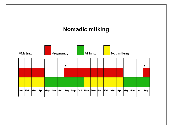
Fig. 1 Traditional
dairy farming practice
Our survey in Mongolia found that cows fed only on pasture do not lactate during the latter half of pregnancy. Mongolian cows become pregnant as a result of natural mating in July or August, and give birth to calves in April or May. The Mongolian nomads milk their cows for 5 months from June to October, obtaining, at best, 5 L milk per day from a cow (Fig. 1). Pregnant cows are milked only in the first trimester of pregnancy. One of these nomads told us "Cows produce milk for the calves of the next generation, not for humans. If we steal too much milk from pregnant cows, they cannot give us good calves".
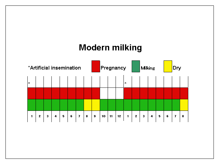
Fig. 2 Modern dairy farming
practice
However, modern genetically-improved dairy cows, such as the Holstein, which are artificially inseminated three months after calving, are usually kept on a combination of grass and concentrates (grain/protein mixes and various by-products). They continue to lactate for almost the whole of pregnancy, extending the milk-producing period to 305 days per year (19) (Fig. 2).
Estrogens in Milk and Dairy Products
There is about 30 pg/mL estrone sulfate in the whey fraction of milk from non-pregnant cows (15). During pregnancy, however, this level becomes elevated to 151pg/mL from day 41 to day 60, and to maximum levels of about 1,000 pg/mL from 220 to 240 days of gestation. According to Gyawu and Pope (12), peak levels of circulating estrogens in cows of late pregnancy are several hundred fold higher than those in non-pregnant cows during ovulation, which suggests that milk from cows in late pregnancy contains much larger amounts of estrogens than does milk from non-pregnant cows.
The oral bioactivity of estradiol and estrone may be relatively low. According to Andersson and Skakkebaek (22), however, estrone sulfate has a high oral activity, and once inside the body it can be converted to estrone and estradiol.
In addition to estrogens, milk contains a large amount of another female hormone, progesterone, at concentrations ranging from 1,400 pg/mL in skim milk to 10,000 pg/mL in whole milk and 300,000 pg/g in butter (14).
Batra et al. (10) measured the levels of estradiol-17b in plasma and whole milk of Murrah buffaloes by radioimmuno assay, and found a very close correlation between levels in plasma and milk; the estradioI concentration in milk was approximately twice that in plasma. Thus, hormone concentrations in whole milk can exceed those in plasma, probably because of synthesis in the mammary gland (11). This phenomenon has been used to diagnose pregnancy in cattle by analyzing the progesterone, estrone, or estradiol-17b content of milk (15,23).
It might be argued that the increased volume of milk per cow per day in modern dairy farming would dilute the estrogens transferred from the circulation of a cow to the milk. However, modern milk contains almost the same amounts of protein, fat, calcium, etc., as traditional milk; it is unlikely that the mammary gland of a high-producing dairy cow secretes nutrients selectively, leaving estrogens alone in the circulation.
Estrogens and Male Reproductive Organs
Endogenous estradiol-17b secreted by the ovaries in the female, exerts negative feedback effects on the secretion of gonadotropins FSH and LH, affecting the hypothalamo-pituitary system (9,24-26). FSH regulates the multiplication of Sertoli cells, which, in turn, are responsible for orchestrating and regulating spermatogenesis. Estrogens can not only impair the development of the male reproductive tract via physiological pathways, but also alter the multiplication of Sertoli cells. In humans, significant qualitative and quantitative changes in the Sertoli cell population take place even after birth, especially during the prepubertal period (27).
Animal studies have shown that alteration of the Serto1i cell number in early life determines testicular size and sperm output in adulthood (28). In rats, the oral dose of exogenous estradioI sufficient to induce sterility was reported to be high (29). However, in the male rat, intraperitoneal doses as low as 10 ng estradiol/rat were reported to induce morphological lesions and spermatogenic arrest without affecting gonadotropin release, after 21 dai1y exposures (30). This suggests that estradiol has a direct effect on developing spermatids.
The dose of 10 ng used for rats (30) can be extrapolated to humans by using body surface area, on the assumption that the two species have the same sensitivity to exogenous estrogens. Assuming that body surface area is proportional to body weight (kg) to the power of 0.7 (31), i.e., (body weight)*0.7, then 10 ng of estradiol in a rat (0.1 kg) corresponds to 410 or 540 ng of estradiol in a child weighing 20 or 30 kg, respectively.
This extrapolation certainly cannot be applied directly to the estrogens in milk, since the bioavailability of estrogens taken orally may differ from that of intraperitoneally administered estrogens (14). However, since no data are available on the species difference in susceptibility to exogenous estrogens, and since susceptibility is expected to differ greatly among children, it is not unreasonable to suggest that several hundred nanograms of estrogens from foods might be enough to affect spermatogenesis in particularly susceptible boys.
Adverse Effects of Milk on Male Reproductive Organs
The adverse effects of milk on male reproductive organs are only sparsely discussed in the literature. One reason may be that precise evaluation of individual milk intake is difficult, because milk and its products (cheese, cream, butter, fermented milk, powdered milk, etc.) are used in a variety of foods, including cakes, candies, ice cream, and chocolates.
Davies et al. (32) tested the hypothesis that milk and dairy products are risk factors for testicular cancer in a case-control study undertaken in East Anglia, UK. All the cases were men with testicular cancer, and for each of the 200 cases, there were four controls: two cancer controls and two population controls. All the responding subjects completed a dietary questionnaire that included questions on their current and adolescent consumption of milk, dairy products, fruits, and vegetables. Cases had consumed significantly more milk in adolescence than controls.
In a case-control study conducted in Northern Italy on 96 histologically confirmed cases and 292 controls with acute, non-neoplastic or genital tract disease, milk was identified as a risk factor for prostate cancer (33). There was a significant increase in risk with frequency of milk consumption: compared with nondrinkers or occasional milk drinkers, the relative risk was 5.0 (95% confidence interval 1.5-16.6) for two or more glasses per day.
Estrogens in Milk and Prepubertal Children
Exposure to exogenous sex steroids is a greater risk for prepubertal children, since their endogenous levels are extremely low (22). Hartmann et al. (14) estimated the average daily intake of animal-derived estrogens (estradiol-17b and estrone) for prepubertal boys to be 80 ng, with milk and dairy products accounting for 60-70% of this. Nonetheless, Hartmann et al. concluded that no hormonal effects could be expected from naturally occurring sex steroids in food, because this value is far exceeded by steroid production in prepubertal boys.
However, the value 6,500 ng/day given by JECFA-WHO (34) for the daily production rate (PR) of estradiol in prepubertal boys is a significant overestimate, according to Andersson and Skakkebaek (22), JECFA-WHO used the metabolic clearance rate for adult women and serum estradiol levels higher than realistic, when calculating the PR in prepubertal boys. Therefore, prepubertal boysš average intake of 80 ng/day of food-derived estrogens (14) may not be negligible.
Milk Consumption in Japan
Sharpe and Skakkebaek (9) stated that consumption of dairy products is too excessive in developed countries, a trend that probably started in the1940s and 1950s.
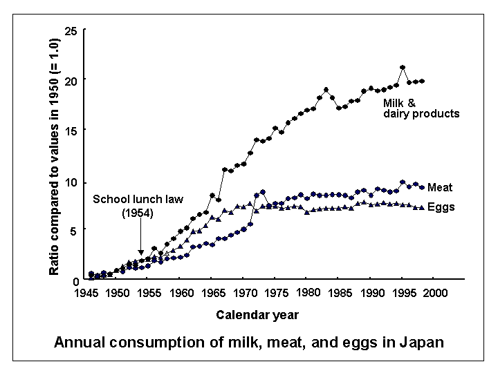
Fig. 3 Consumption of animal products in Japan
This holds true in Japan, where a no-meat/no-milk culture essentially prevailed until the end of World War Two. In the past 50 years, Western dishes using more animal products as ingredients have become popular in Japan (Fig. 3) (35).
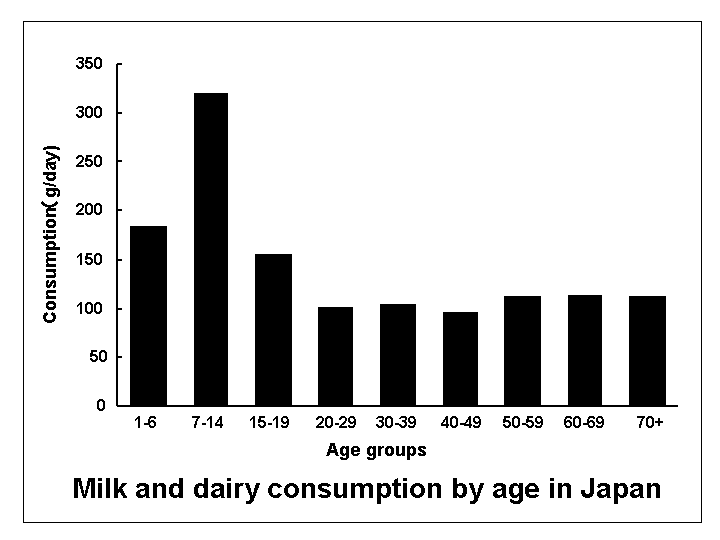
Fig. 4 Milk consumption
of Japanese prepubertal boys
Each Japanese child is given 200 mL of milk daily at lunch in school. For this reason, the average daily milk consumption of this age group reaches 322 g (36), a remarkable quantity in Japan, where the population's average milk consumption is as low as 135 g/day (Fig. 4). Pregnant women in Japan are also encouraged to consume milk and dairy products, to meet their calcium requirements during pregnancy.
Commercial milk in Japan contained estrone 21.8-26.0 pg/mL, estradiol-17b 25.1-33.6 pg/mL, and estriol 31.4-98.6 pg/mL (authorsš unpublished data). By drinking 300 mL of milk per day, a childšs intake of estradiol-17b alone, the most potent estrogenic hormone, is approximately 10 ng per day. This is 4,000 times the intake of environmental hormones of xenoestrogens, in terms of hormone activity (37).

Fig.
5 Death rate of prpstate cancer in Japan
Between1950 and 1997 in Japan, milk consumption per person per day increased almost 20-fold, from less than 7 g to 135 g (Fig. 3). This sudden increase in consumption of estrogenic milk might have affected the reproductive organs of Japanese males. For example, the incidence rate of prostate cancer in Japan is increasing at the fastest rate in the world (38), and the associated age-adjusted death rate has risen almost 13-fold during the last 45 years, from 0.45/100,000 in 1950 to 5.93/100,000 in 1995 (Fig. 5).
Reproductive health should be monitored carefully in Japan, where milk consumption started to increase only 40 years ago, and where prepubertal children are the major consumers. Epidemiological studies into reproductive outcomes in Japan will certainly shed light on the relationship between milk and male reproductive health.
Conclusion
The increased incidence of male reproductive disorders over the last 50 years has attracted much attention. Although great concern has been raised over the role of environmental compounds with estrogenic activity in the development of hormone-related diseases, exposure to exogenous sex steroids from food has not been widely discussed. Humans consume milk from cows in the latter half of pregnancy, when estrogen levels are markedly elevated. Estradiol-17b is about 10,000-fold more biopotent than most identified environmental xenoestrogens (22). For prepubertal boys in the critical period of sexual development, excess consumption of milk and dairy products might interfere with the physiological development of the reproductive system and result in decreased semen quality in adulthood. Epidemiological or experimental studies should be undertaken to test our hypothesis that milk is responsible, at least in part, for male reproductive disorders.
References
1. Auger J,
Kunstmann JM, Czyglik F, Jouannet P. Decline in semen quality among fertile
men in Paris during the past 20 years. N Engl J Med 1995; 332: 281-285.
2. Carlsen E, Giwercman A, Keiding N, Skakkebaek NE. Evidence for
decreasing quality of semen during past 50 years. BMJ 1992; 305: 609-613.
3. Carlsen E, Giwercman A, Skakkebaek NE. Declining sperm counts
and increasing incidence of testicular cancer and other gonadal disorders:
is there a connection? Irish Med J 1993; 86: 85-86.
4. Irvine S, Cawood E, Richardson D, MacDonald E, Aitken J. Evidence
of deteriorating semen quality in the United Kingdom: birth cohort study
in 577 men in Scotland over 11 years. BMJ 1996; 312: 467-471.
5. Bergstro R, Adami HO, Moner M, Zatonski W, Storm H, Ekbom A,
Tretli S. Teppo L, Akre O, Hakulinen T. Increase in testicular cancer
incidence in six European countries: a birth cohort phenomenon. J Natl
Cancer Inst 1996; 88: 727-733.
6. McKiernan JM, Goluboff ET, Liberson GL, Golden R, Fisch H. Rising
risk of testicular cancer by birth cohort in the United States from 1973
to 1995. J Urol 1999; 162: 361-363.
7. Pajarinen J, Laippala P, Penttila A, Karhunen PJ. Incidence
of disorders of spermatogenesis in middle aged Finnish men, 1981-91: two
necropsy series. BMJ 1997; 314: 13-18.
8. Swan SH, Elkin EP, Fenster L. Have sperm densities declined?
A reanalysis of global trend data. Environ Health Perspect 1997; 105:
1228-1232.
9. Sharpe RM, Skakkebaek NE. Are oestrogens involved in falling
sperm counts and disorders of the male reproductive tract? Lancet 1993;
341: 1392-1395.
10. Batra SK, Arora RC, Bachlaus NK, Pahwa GS, Pandey RS. Quantitative
relationships between oestradiol-17beta in the milk and blood of lactating
buffaloes. J Endocrinol 1980; 84: 205-209.
11. Erb RE, Chew BP. Keller HF. Relative concentrations of estrogen
and progesterone in milk and blood, and excretion of estrogen in urine.
J Animal Sci 1977; 45: 617-626.
12. Gyawu P, Pope GS. Oestrogens in milk. J Steroid Biochem 1983;
19: 877-882.
13. Hamon M. Fleet IR. Heap RB. Comparison of oestrone sulphate
concentrations in mammary secretions during lactogenesis and lactation
in dairy ruminants. J Dairy Res 1990; 57: 419-422.
14. Hartmann S, Lacorn M, Steinhart H. Natural occurrence of steroid
hormones in food. Food Chem 1998; 62: 7-20.
15. Heap RB, Hamon M. Oestrone sulphate in milk as an indicator
of a viable conceptus in cows. Br Vet J 1979; 135: 355-63.
16. Pope GS, Swinburne JK. Reviews of the progress of dairy science:
hormones in milk: their physiological significance and value as diagnostic
aids. J Dairy Res 1980; 47: 427-449.
17. Gurr MI. Male sexual development in ęa sea of oestrogenė. Lancet
1993; 342: 125-126.
18. Hurt RD. American Agriculture: A Brief History. p 53, Iowa
State University Press, Ames, 1994.
19. Japan Dairy Farming. http://jdc.lin.go.jp/dairy/farm13.htm,
2000.
20. Frink CR, Waggoner PE, Ausubel JH. Nitrogen fertilizer: retrospect
and prospect. Proc Natl Acad Sci USA 1999; 96: 1175-1180.
21. Brown LR. Seeds of Change. The Green Revolution and Development
in the 1970šs. pp 3-12, Praeger Publishers, New York ß Washington ß London,
1970.
22. Andersson AM, Skakkebaek NE. Exposure to exogenous estrogens
in food: possible impact on human development and health. Eur J Endocrinol
1999; 140: 477-485.
23. Hamon M, Fleet IR, Holdsworth RJ, Heap RB. The time of detection
of oestrone sulphate in milk and the diagnosis of pregnancy in cows. Br
Veter J 1981; 137: 71-77.
24. Bellido C, Pinilla L, Aguilar R, Gaytan F, Aguilar E. Possible
role of changes in post-natal gonadotrophin concentrations on permanent
impairment of the reproductive system in neonatally oestrogenized male
rats. J Reprod Fertil 1990; 90: 369-374.
25. Gaytan F, Pinilla L, Aguilar R, Lucena MC, Paniagua R. Effects
of neonatal estrogen administration on rat testis development with particular
reference to Sertoli cells. J Androl 1986; 7: 112-121.
26. Sharpe RM, Atanassova N, McKinnell C, Parte P, Turner KJ, Fisher
JS, Kerr JB, Groome NP, Macpherson S, Millar MR, Saunders PT. Abnormalities
in functional development of the Sertoli cells in rats treated neonatally
with diethylstilbestrol: a possible role for estrogens in Sertoli cell
development. Biol Reprod 1998; 59: 1084-1094.
27. Cortes D, Muller J, Skakkebaek NE. Proliferation of Sertoli
cells during development of the human testis assessed by stereological
methods. Int J Androl 1987; 10: 589-596.
28. Orth JM, Gunsalus GL. Lamperti AA. Evidence from Sertoli cell-depleted
rats indicates that spermatid number in adults depends on numbers of Sertoli
cells produced during perinatal development. Endocrinology 1988; 122:
787-794.
29. Biegel LB, Flaws JA, Hirshfield AN, OšConnor JC, Elliott GS,
Ladics GS, Silbergeld EK, Van Pelt CS, Hurtt ME, Cook JC, Frame SR. 90-Day
feeding and one-generation reproduction study in Crl:CD BR rats with 17b-estradiol.
Toxicol Sci 1998; 44: 116-142.
30. Seegers JC, van Aswegen CH, Nieuwoudt BL, Joubert WS. Morphological
effects of the catecholestrogens on cells of the seminiferous tubules
of Sprague-Dawley rats. Andrologia 1991; 23: 339-345.
31. Adolph EE. Quantitative relations in the physiological constitutions
of mammals. Science 1949; 109: 579-585.
32. Davies TW. Palmer CR. Ruja E. Lipscombe JM. Adolescent milk,
dairy product and fruit consumption and testicular cancer. Br J Cancer
1996; 74: 657-660.
33. La Vecchia C, Negri E, DšAvanzo B, Franceschi S, Boyle P. Dairy
products and the risk of prostatic cancer. Oncology 1991; 48: 406-410.
34. JECFA (The Joint FAO/WHO Expert Committee on Food Additives)-WHO.
Evaluation of certain veterinary drugs residues in food. Thirty-second
Report of the Joint FAO/WHO Expert Committee on Food Additives. WHO Technical
Report Series 763, WHO, Geneva, 1988.
35. Health Promotion and Nutrition Division, Health Service Bureau,
Ministry of Health and Welfare of Japan. The National Nutrition Survey
in Japan, 1948-1999, Dai-ichi Shuppan, Tokyo (in Japanese).
36. Health Promotion and Nutrition Division, Health Service Bureau,
Ministry of Health and Welfare of Japan. The National Nutrition Survey
in Japan, 1999, Dai-ichi Shuppan, Tokyo (in Japanese).
37. Safe SH. Environmental and dietary estrogens and human health:
is there a problem? Environ Health Perspect 1995; 103: 346-351.
38. Hsing AW, Tsao L, Devesa SS. International trends and patterns
of prostate cancer incidence and mortality. Int J Cancer 2000; 85: 60-67.
ü@